Each skeletal muscle has three layers of connective tissue that enclose it provide structure to the muscle and compartmentalize the muscle fibers within the muscle. Sarcomere Lower middle box Step-by-step explanation A band The thick myosin filaments are arranged across the myofibrils and the cell causing them to refract light and generate the A Band a dark band.

Sarcomere Structure Anatomy And Physiology Physiology Knowledge
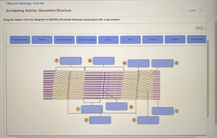
Reset Help A Band Barmere Hand Band MI Art-Labeling Activity.
. Sarcomere structure The Structure Of A Skeletal Muscle Fiber Part A Drag The Labels Onto. Art labeling are drag-and-drop activities that allow students to assess their knowledge of terms and structures as well as the order of steps and elements involved in physiological processes. The Structure Of A Skeletal Muscle Fiber Part A Drag The Labels Onto The Diagram To Identity Structural Features Associated With A Skeletal Muscle Fiber.
The structure of a skeletal muscle fiber Part A Drag the labels onto the diagram to identity structural features associated with a skeletal muscle fiber. Sarcomere Structure Drag the labels onto the diagram to identity structural features associated with a sarcomere. This would also be a great way to review for a.
Layer B is composed primarily of _____. Art labeling sarcomere structure art-labeling activity. SUNY Buffalo State College BIO 308.
Art Labeling Activity Antibody Structure 13 Of 14 Drag The Labels To Identify The Structural Components Of An Antibody React Help Heavy Chain Stofbinding To Macrophages Light Chain Anogen Biding Site Of Light Read the latest research on diseases and conditions symptoms new treatment options and more. Structural organization of skeletal muscle Reset Help Epimysium Muscle fascicle Endomysium Perimysium Nerve Muscle fibers Blood vessels Tendon Muscle fiber cell. Diagram and label a sarcomere including a thick filament thin filament A band H zone I band Z A.
However thick and thin filamentsthe components of sarcomeresdo not shorten. Skin that has four layers of cells is referred to as thin skin From deep to superficial these layers are the. Solved Art Labeling Activity Sarcomere Structure Drag The Chegg Com The A band stays the same.
Pages 80 Ratings 81 21 17 out of 21 people found this document helpful. Start studying the Art- labeling Activity flashcards containing study terms like and more. Review sarcomere structures and the components of muscle contraction with this informal assessmenthomeworkactivity.
Memorize flashcards and build a practice test to quiz yourself before your exam. Structure of the epidermis PartA Drag the appropriate labels to their respective targets. Vi skulle vilja visa dig en beskrivning här men.
Students are directed to draw label and color code their model of a sarcomere as well as muscle contraction. Reset Help A band bend Thinaments Mline Thick framanta Sarcomero Zine Tion Zone of overlap O Subna Request Answer Posted 4 months ago. Ooo baby I love your way.
These tissues include the skeletal muscle fibers blood vessels nerve fibers and connective tissue. Small Intestine Art-labeling Activity. Label the parts of the Sarcomere flashcards containing study terms like Z- line Actin thin filaments Myosin thick filaments and more.
A simple activity about the digestive system that could be used to introduce the topic as a bell ringer activity or as an assessment at the end of the topicWord document format so can be modified to suit your needsThere are two activities included-. Reset Help A band Barmere Hand band MI Art-labeling Activity. Art labeling activity the structure of a skeletal.
There is a bright region between the A bands with no thick myofilaments and just thin actin filaments. Match to correct box. Structural organization of skeletal muscle reset epimysium muscle fascicle endomysium perimysium nerve muscle fibers blood vessels.
This will allow students to get creative and master the muscle content. The Structure of a Sarcomere Part A Drag the labels to the appropriate location in the figure. The structure of a skeletal muscle fiber part a drag the.
Reset Help Thick lament Thin filament Elastic ftamenti 1 band Mlino H zone Sarcomero Z disc Zone of overlap A band CCD WWW. The structure indicated by label E is part of which of the following. Course Title AA 1.
The Structure Of A Sarcomere Part A Drag The Labels To The Appropriate Location In The Figure. 1-countour drawing of human body head - drawn on bottom paper. Each skeletal muscle is an organ that consists of various integrated tissues.
Figure as an art labeling activity. Structure and Bands of the Sarcomere Drag the appropriate labels to their respective targets. Antibody Structure Drag The Labels To Identify The Structural.
Art-Labeling and Art-Based Questions give students practice identifying structures and process steps using art from the book. Start studying Art-labeling Activity. Nail Structure - Art Labeling Activity Vertebral Anatomy Jpg M No Subject Stellarvore91 Gm X G Anatomy Abdominal Quadrants X Course Home X Smart Pearson Course Hero - Reset help nail root of nai plate nail body of nail plate prodmal nail fold hyponychium lateral nail foid eponychium nail bed lunula nail matri medial nail fold free adge.
The struc Written By stele Saturday June 4 2022 Add Comment bob i love wallpaper ooh baby i love your way bob marley Everyday yeah yeah I wanna be with you night and day. This preview shows page 7 - 11 out of 80 pages. Students who viewed this also studied.

Art Labeling Activity Sarcomere Structure Diagram Quizlet
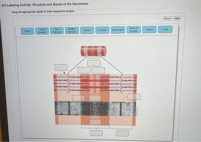
Solved Art Labeling Activity Structure And Bands Of The Chegg Com

Solved Art Labeling Activity Sarcomere Structure Drag The Chegg Com
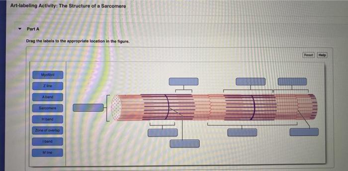
Solved Art Labeling Activity The Structure Of A Sarcomere Chegg Com

Art Labeling Activity The Structure Of A Skeletal Muscle Fiber Diagram Quizlet

Label The Sarcomere Structure Diagram Quizlet
Solved Muscle Histology Post Lab Art Labeling Activity Chegg Com
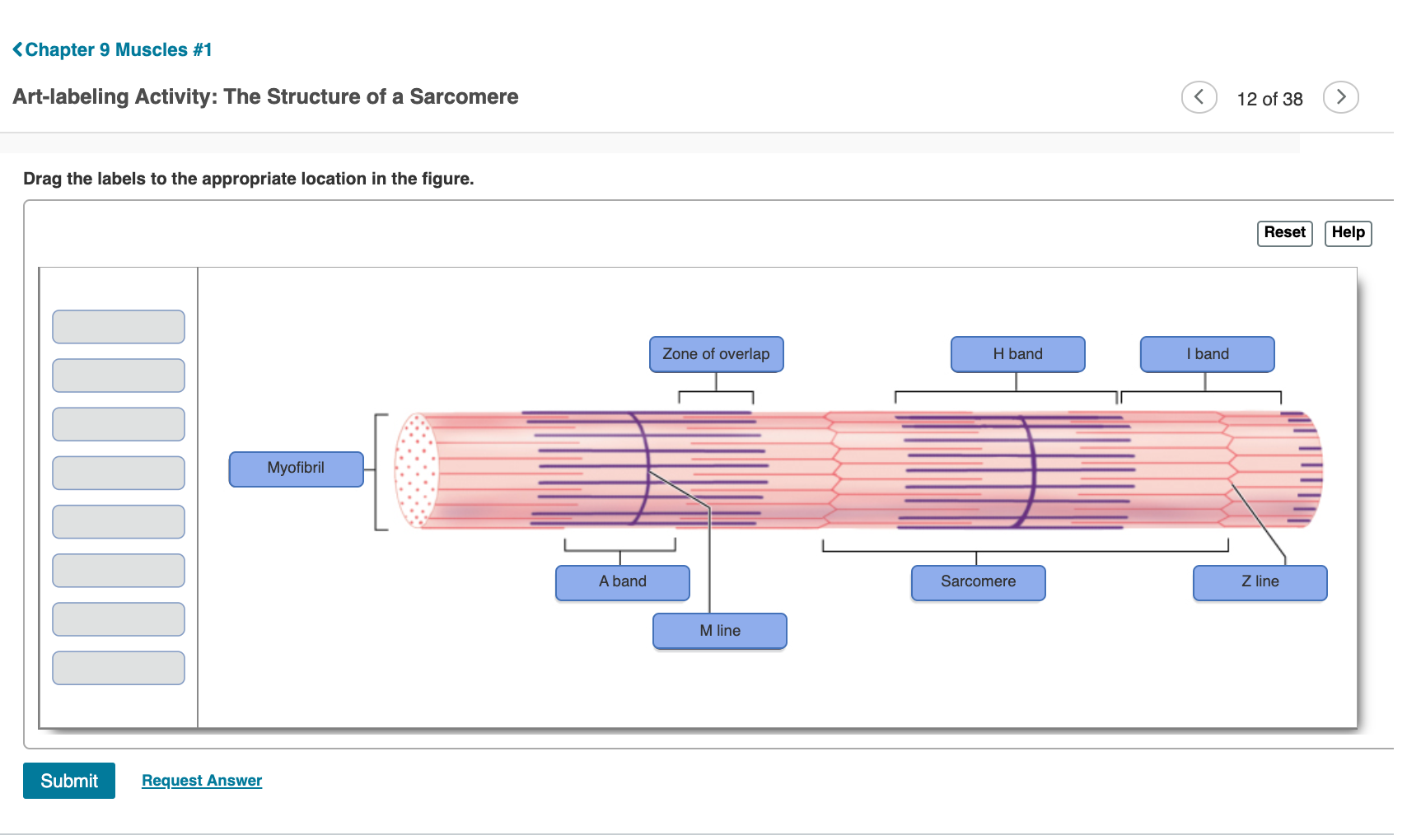
Solved Chapter 9 Muscles 1 Art Labeling Activity The Chegg Com
0 comments
Post a Comment